3d atlas mouse anatomy
For more information please refer to the documentation. In this thesis I describe how to construct a high-resolution 3D atlas of the mouse brain from 2D microscopic images.

Hypothalamus Anatomy Physiology Wikivet English Anatomy And Physiology Physiology Dog Anatomy
Ng and colleagues broke up the.

. Successful integration of these data requires a standard 3D reference atlas. Zygote Body is a free online 3D anatomy atlas. Anatomy 3D Atlas allows you to study human anatomy in an easy and interactive way.
Previous versions of the atlas were lower resolution 3D maps while CCFv3s resolution is fine enough that it can pinpoint individual cells locations. The critical factors are alignment of section images and matching of stain intensities because their precision controls the efficiency and accuracy of 3D segmentation. Hold right click while dragging your mouse across the screen.
The Mouse Limb Anatomy Atlas provides a novel and valuable tool for researchers studying limb development and can be applied to a range of research areas including the identification of abnormal limb patterning in transgenic lines and studies of models of congenital limb abnormalities. Ad Browse discover thousands of brands. It can take you through cross-sections of the human and mouse brain and also maps out genes across each brain region in the mouse brain.
In 2011 the reference atlas was updated to enable interactive online exploration of the atlas and to provide a deeper level of 3-D annotation for informatics analysis and viewing in the Brain Explorer 3-D viewer. In addition the atlas demonstrates. To plot such detailed anatomy manually would be an.
View isolate and learn human anatomy structures with Zygote Body. March 21th 2000 By. Ad The worlds largest software App discovery destination.
This is the best way to see the anatomy of the brain in 3D. The Allen Brain Atlas was created by the Allen Institute for Brain Science. Free shipping on qualified orders.
You can filter for gene expression and compare across the thousands of. Recent large-scale collaborations are generating major surveys of cell types and connections in the mouse brain collecting large amounts of data across modalities spatial scales and brain areas. The large sample size of 35 mouse embryos coupled with the acquisition of 28 μm 3 resolution 3D micro-CT images and the manual segmentation of a total of 48 anatomical structures 25 within the brain alone results in the most comprehensive mouse embryo representative average atlas in the literature.
Ad 1Free Stock Images2Royalty Free Photos3Clip Art4Backgrounds5Vectors. The complete Skeletal System and a few other contents are always freely accessible enabling you to try the app properly. Complete Cranial Cerebrovascular Anatomy with Neck Vasculature 1a as well as Arterial 1b Anatomy AloneThese models provide an overview rendition of the complete 3D venous and arterial cerebrovascular tree.
Here we present the Alle. Place two fingers on the screen and move them together in the same direction. Press down with two fingers and drag together.
Another immediate goal is to create a 3D voxel atlas of the C57BL6J mouse brain from the present 2D section images. Use the model select icon above the anatomy slider on the left to load different models. LINK TO THIS STEP.
The Home button resets the view. Read customer reviews find best sellers. Code may be re-used for non-commercial use.
While 3D imaging methods such as MRI and CT have been widely applied 2D imaging methods such as optical microscopy typically generate images with much higher resolution. Reconstructed to make 3D models and annotated using. 132 coronal sections evenly spaced at 100 µm intervals and annotated to.
Discover Over 400000000 Royalty-Free Images Plus 150000 New Added Daily. Plays an important role in modern biology and medical science. This version is based on the atlashtml program.
Free easy returns on millions of items. Alternatively hold shift while clicking and dragging across the screen. To make the atlas Dr.
All soft tissue bony and neural elements have been removedThe structures of the arterial and venous systems have been. This app is freely downloadable however in-app purchase is required to unlock the contents. The Best downloads for any device.
Edmund Cape Last updated. Or use the buttons in the upper left. The CCFv3 atlas builds on a partial version released in 2016 that mapped the entire mouse cortex the outermost shell of the brain.
Clickdrag with the mouse to rotate scroll to zoom.

Pin Page

Pin On Ibclc Study Aids

Dynamic Networks In The Emotional Brain Pubmed

Pin Page

Anatomy Drawing Hand Video Figur Cizimleri Cizim Cizimler

Anatomical Basis Of Wallenberg Lateral Medullary Syndrome Mnemonic

Animal Communication Web Topic 10 4 Brain Diagram Animal Communication Limbic System

Ontogenetic Scaling Patterns And Functional Anatomy Of The Pelvic Limb Musculature In Emus Dromaius Novaehollandiae Leg Muscles Anatomy Muscle Anatomy Anatomy

Tiny Treasures Award Winning Microscopic Pics

Mouse Brain Atlas

Earth Globe 3d Render Of An Earth Globe Ad Globe Earth Render Globe Earth Ad Earth Globe Illustration Stock Illustration

Allen Institute For Brain Science Announces Mapping Of The Mouse Cortex In 3d

Brain Nonneoplastic Lesion Atlas
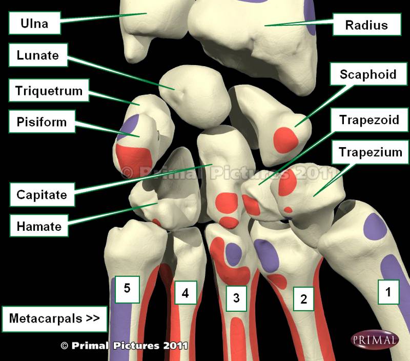
ป กพ นในบอร ด Bones Hand
Pin Page

Unprecedented 3 D Images Of Human Ear Anatomy For Hearing Restoration The New 3d Images Of The Ear Provide Informatio Human Ear Anatomy Ear Anatomy Human Ear

Postmortem Examination Of Patient H M S Brain Based On Histological Sectioning And Digital 3d Reconstruction Nature Communications

3d Reconstruction Of Neurons

Brainbow